Researchers from NAIST have developed a machine learning-based method for the opportunistic screening and early diagnosis of bone conditions
Osteoporosis is a prevalent medical condition characterized by low bone mineral density (BMD), resulting in weakened and brittle bones. Individuals with osteoporosis face an elevated risk of fractures and have reduced daily functionality. The gold standards for diagnosing osteoporosis include dual-energy X-ray absorptiometry (DXA) and quantitative computed tomography (QCT), but these methods require specialized equipment and can be expensive. Consequently, more accessible and cost-effective modalities for osteoporosis screening are warranted. While machine learning methods to estimate BMD from X-ray images have recently gained impetus, they often require large training datasets.
Addressing this problem, researchers from Japan have now developed a novel method that uses a machine learning approach called the hierarchical learning framework to estimate BMD from plain X-ray images. The research group--led by Yi Gu, Yoshito Otake, Yoshinobu Sato from Nara Institute of Science and Technology (NAIST) and Keisuke Uemura from Osaka University--published their findings in the journal Medical Image Analysis.
Otake explains the motivation behind this study: "Osteoporosis is generally diagnosed at advanced stages once its symptoms become apparent. X-ray images can be valuable for opportunistic diagnosis, but efficiently extracting BMD information from these has been a significant challenge. We hoped to solve this problem by using information derived from the computed tomography (CT) image in the training stage to develop a model for an accurate, efficient, and explainable BMD estimation solely from an X-ray image."
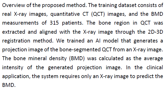
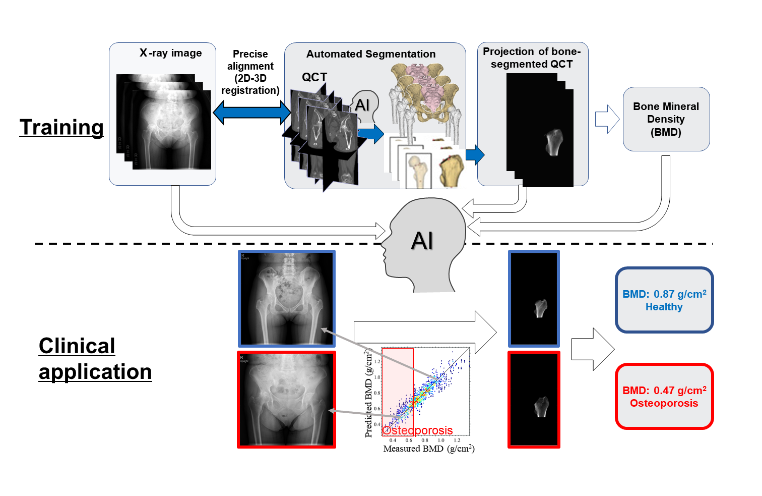
To create the training dataset, the virtual X-ray image of the bone region that was precisely aligned with the patient's X-ray image was first reconstructed using original quantitative CT (QCT) scans from patients. Subsequently, three training steps were performed to develop the final BMD estimation model. In the first and second stages, X-ray images were decomposed into the virtual X-ray image of the bone region. In the third stage, the model learned the correlations between the virtual X-ray images and BMD values based on the training dataset. As a result, it could estimate BMD using a single X-ray image. Importantly, this approach led to high performance with a couple of hundred datasets of CT and X-ray image pairs. In addition to the BMD value, i.e., the average bone density within a pre-defined region, the model produces the virtual X-ray image of the QCT of the bone region representing the distribution of the bone density, significantly enhancing the explainability of the prediction result.
To understand if this model could serve as an alternative to gold standard methods such as DXA and QCT, validation studies were performed using real clinical datasets. BMD values obtained using the proposed method showed 0.88 and 0.92 correlation with those obtained using DXA and QCT, respectively, highlighting its reliability. Further validation experiments showed that this novel method was also highly robust. It allowed for consistent BMD estimation irrespective of variations in patient poses and the level of image compression.
Altogether, the results are a testament to the immense potential this newly developed method has in routine clinical practice. Excited about its prospects, Otake comments, "Our proposed method can increase the accessibility of BMD estimation; it can be used anywhere, including centers that do not have sophisticated equipment for BMD measurement."
Indeed, the method could allow for opportunistic screening and detailed checkups in the treatment follow-up in clinical practice, enabling early diagnosis and intervention and improved quality of life for patients with osteoporosis.
###
Resource
- Title: Bone mineral density estimation from a plain X-ray image by learning decomposition into projections of bone-segmented computed tomography
- Authors: Yi Gu, Yoshito Otake, Keisuke Uemura, Mazen Soufi, Masaki Takao, Hugues Talbot, Seiji Okada, Nobuhiko Sugano, Yoshinobu Sato
- Journal: Medical Image Analysis
- DOI: 10.1016/j.media.2023.102970
- Information about the Imaging-based Computational Biomedicine Laboratory can be found at the following website: https://isw3.naist.jp/Research/ai-icb-en.html