
Researchers at Nara Institute of Science and Technology create a lab-on-a-chip with a viscoelastic fluid that can separate spherical from elongated bacteria, which can help standardize biological research and improve medical testing
Scientists at Nara Institute of Science and Technology (NAIST) achieved shape-based separation of Escherichia coli (E. coli) bacteria using a viscoelastic fluid inside microfluidic channels. This "lab-on-a-chip" system has the capability to assist in making scientific experiments more reproducible, as well as provide more accurate assessments of the severity of bacterial infections based on patient samples.
The task of separating tiny bacteria based on their shape is a vital step in scientific and clinical applications, but also a difficult one. Whether an E. Coli bacterium is in a round or elongated configuration can indicate its state of biological function, and the ability to prepare homogeneous populations with uniform shapes may help improve experimental reproducibility. Moreover, being able to sort samples into sub-populations based on their shape may help diagnose patient health or assess environmental contamination. However, this ability has been difficult to achieve, especially at the scales needed to be practical.
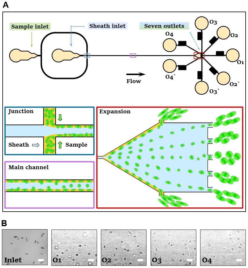
Now, a team of researchers at NAIST and has been able to separate drug-treated E. coli bacteria using microfluidic channel containing poly-ethylene-oxide (PEO), a viscoelastic fluid. The elongation of the bacteria, as measured by the aspect ratio, ranged from 1.0 (spherical) to 5.5 (rod-shaped). Microchannels made of poly(dimethylsiloxane) were created with photolithography. PEO, which has both high viscosity and elasticity, was made to flow along the edges of an expanding V-shaped chamber. The elongated bacterium followed this flow to be collected at the edges of the device. "The shape of a E. coli bacterium changes based on its attachment, motility, dispersal, predation and differentiation behaviors," author Yalikun Yaxiaer explains. A rod-like configuration is more efficient for swimming, while a spherical shape may help it form communities. These changes are important milestones for the progression of disease, and whether the bacteria are swimming free or forming communities. "This technology will allow downstream genomics experiments on the separated E. coli populations to determine linkage between cell shape, antibiotic susceptibility and gene function," author Yoichiroh Hosokawa says. The team hopes the microfluidic platform they developed will be adopted by the broader scientific community for different applications in the fields of microbiology, molecular biology and biomedicine.
###
Resource
- Title: Shape-based separation of drug-treated Escherichia coli using viscoelastic microfluidics
- Authors: Tianlong Zhang, Hangrui Liu, Kazunori Okano, Tao Tang, Kazuki Inoue, Yoichi Yamazaki, Hironari Kamikubo, Amy K Cain, Yo Tanaka, David Inglis, Yoichiroh Hosokawa, Yalikun Yaxiaer & Ming Li
- Journal: Lab on a Chip
- DOI: 10.1039/D2LC00339B
- Information about the Bio-Process Engineering Laboratory can be found at the following
website: https://mswebs.naist.jp/english/courses/list/labo_11.html