

NEWS RELEASE 25-JUN-2026
Peer reviewed publication
NARA INSITUTE OF SCIENCE AND TECHNOLOGY
Self-Propelled Actin Filaments: Novel Structures Driving Spontaneous Cell Morphogenesis
--A Key to Understanding How Cell Shape Emerges Spontaneously--
Summary
Cells possess a remarkable ability for "self-organization" in that they can shape themselves autonomously without external instructions. This phenomenon is fundamental to vital life activities, yet its underlying mechanism remains one of the most significant unsolved mysteries in modern biology.
A research group led by Professor Naoyuki Inagaki, along with Dr. Kio Yagami, Assistant Professor Takunori Minegishi, Assistant Professor Kentarou Baba, Mr. Shinji Misu, Dr. Hiroko Katsuno-Kambe, Dr. Kazunori Okano, Professor Yuichi Sakumura, and Professor Yoichiroh Hosokawa, all from Nara Institute of Science and Technology, Japan, discovered an actin cytoskeleton that actively "runs" within cells, naming it the Self-propelled Treadmilling Actin filament (SpTA). Intriguingly, SpTA moves throughout the cell randomly changing its direction. When an SpTA collides with the cell membrane, it pushes the membrane outward, forming a small protrusion. More SpTAs then accumulate further at this site, driving the growth of the protrusion and ultimately determining the cell's overall shape.
This study illuminates the beginning of the process by which cells generate their form, shedding light on the mechanics of spontaneous cell shaping. Furthermore, the behavior of SpTA closely mirrors that of "self-propelled particles" currently studied in modern physics. This finding is expected to bridge the gap between modern biology and physics, helping us solve the enduring puzzle of self-organization.
Background and Purpose
The cells that make up our bodies change shape with astonishing dynamism. For example, white blood cells--the main players of our immune system--extend foot-like protrusions to migrate and spread hand-like protrusions to engulf foreign invaders such as viruses and bacteria. Similarly, neurons in the brain extend extremely long projections in order to connect with one another and form complex information networks. This inherent ability to generate shape is fundamental to the survival of life.
The cytoskeleton, composed of a protein called actin, supports this morphogenesis. It acts as a structural framework that pushes against the cell membrane from the inside, thereby changing the cell's shape. Previous studies have shown that the actin cytoskeleton is regulated by receiving molecular signals from outside the cell -- a "passive" mechanism driven by external instructions. However, cells can autonomously assemble this framework and create distinct shapes even in the absence of external cues. But how do cells determine their own shape? This "spontaneous" self-organizing mechanism has long remained a mystery, standing as one of the most critical challenges in modern biology.
The research group discovered an actin cytoskeleton that actively runs within cells. Previously, this type of actin cytoskeleton was thought to propagate through the cell as a chemical reaction "wave" and was referred to as an "actin wave". However, this newly observed cytoskeleton does not move like a wave. Instead, it behaves like a "particle" that moves forward, changing direction randomly. This behavior mirrors that of self-propelled particles studied in physics. Consequently, the research group named this entity the Self-propelled Treadmilling Actin filament (SpTA) and investigated its intracellular functions. Traditional physics (equilibrium statistical mechanics) has analyzed particles exhibiting "Brownian motion". In contrast, self-propelled particles can take energy from their environment and convert it into directed motion to propel themselves, and their trajectories involve random fluctuations.
Results
Human glioma U251 cells spontaneously establish their front and rear sides to migrate, even without directional external signals (arrow, Figure 1).
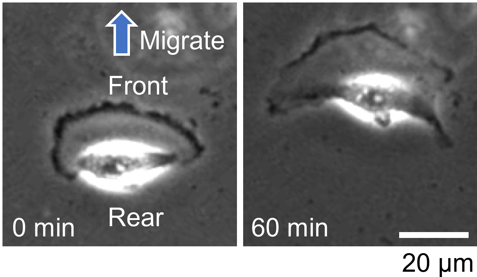
Figure 1. Movement of U251 cells
Using high-resolution live-cell microscopy of the actin cytoskeleton within U251 cells, the research group discovered SpTA actively running inside the cells (Figure 2). SpTA moves throughout the cell while changing directions randomly (arrowheads, Figure 2A). When SpTA collides with the cell membrane, it pushes the membrane outwards to form a small protrusion (arrowheads, Figure 2B).
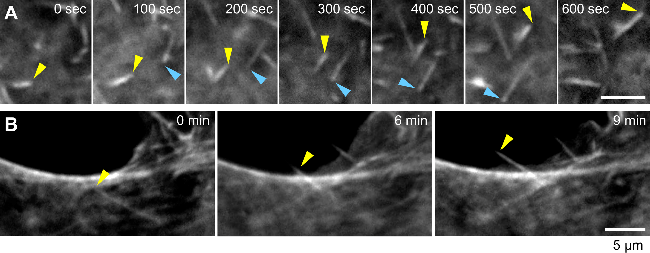
Figure 2. SpTAs running within cells
The team revealed its propulsion mechanism: SpTA utilizes ATP energy to undergo directional polymerization and depolymerization (treadmilling) of actin molecules, thereby advancing through the intracellular space (Figure 3).
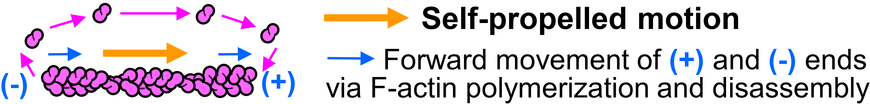
Figure 3. propulsion mechanism of SpTA
Interestingly, SpTAs were also observed accumulating inside these micro-protrusions. To investigate this further, the researchers used laser-fabricated culture dishes to constrain U251 cells into triangular shapes. They found that SpTAs preferentially accumulated at the corners of the triangles (Figure 4A). Furthermore, when the researchers modeled SpTA motility mathematically and ran computer simulations, they obtained identical results (Figure 4B).
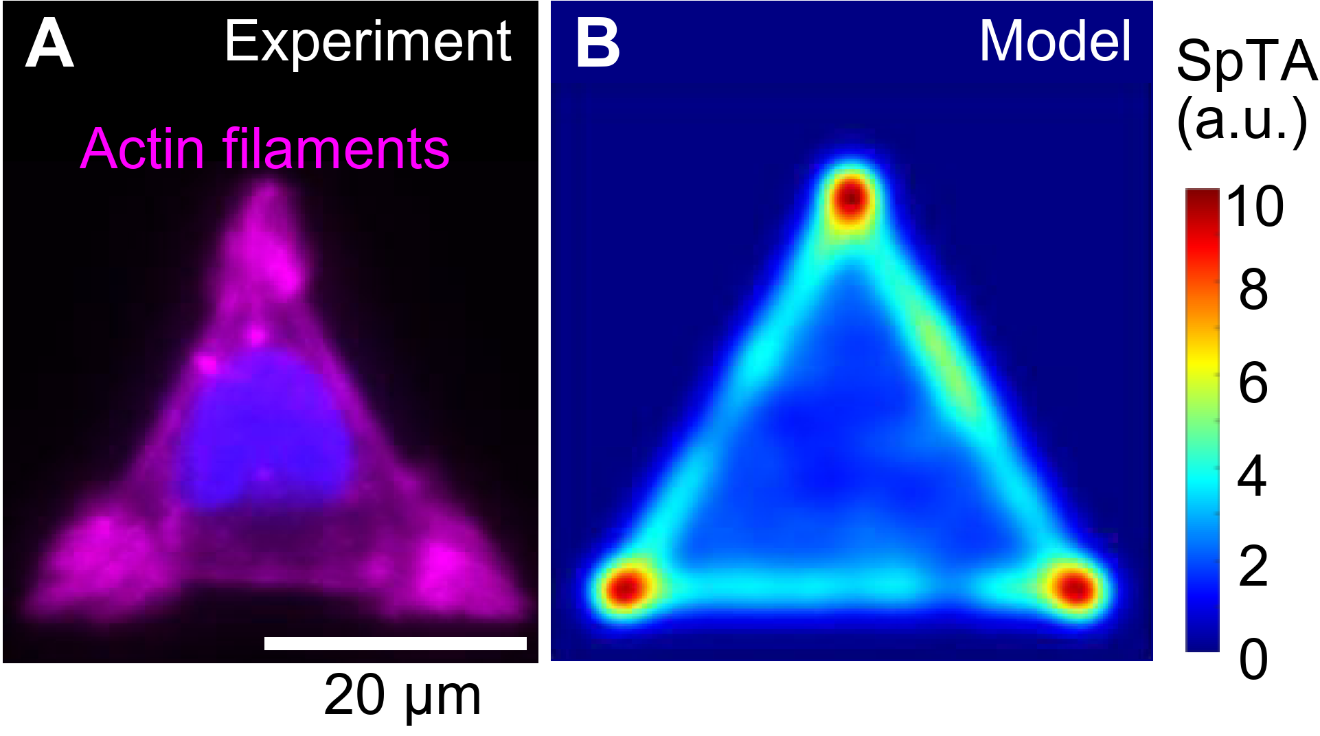
Figure 4. SpTA accumulates at intracellular protrusive regions
Previous physics literature has previously reported that self-propelled particles tend to get trapped in protruding boundaries. Therefore, SpTA possesses an intrinsic property of self-propelled particles to accumulate in protruding regions (i.e., cellular protrusions).

Figure 5. Blocking of SpTA inhibits cell shaping
When the movement of SpTA was suppressed, the formation of the front and rear sides in U251 cells was inhibited (Figure 5), with normal cell migration being disrupted. These findings suggest that SpTA initiates the formation of small protrusions by colliding with and pushing the cell membrane from the inside (Figure 6). The subsequent accumulation of more SpTAs at these sites drives the growth of the protrusions, ultimately generating the overall cell shape (Figure 6).
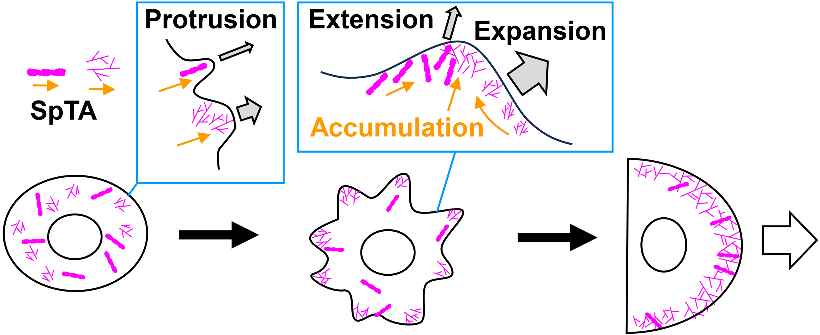
Figure 6. How SpTs drive spontaneous cell morphogenesis
Conclusion
The research group has discovered SpTA, a novel self-propelled particle composed of actin filament assemblies. This study demonstrates a clear multiscale link, whereby microscopic phenomena--the running of SpTA--collectively trigger macroscopic phenomena, such as cell shaping and migration. This insight could help to solve the mystery of biological self-organization. Furthermore, by identifying the self-propelled particle at the origin of self-organization, this study is expected to foster collaboration between modern biology and physics in the study of self-organization.
Resource
Title: Spontaneous membrane protrusion and cell morphogenesis via self-propelled actin filaments
Authors: Kio Yagami, Takunori Minegishi, Kentarou Baba, Shinji Misu, Hiroko Katsuno-Kambe, Kazunori Okano, Yuichi Sakumura, Yoichiroh Hosokawa, and Naoyuki Inagaki
Journal: EMBO Reports
DOI: 10.1038/s44319-026-00804-6
Information about the Laboratory of Systems Neurobiology and Medicine can be found at the following website: https://www.naist.jp/iri/inagaki/english/
Funding information
This research was supported in part by AMED under Grant Number JP17gm0810011, JSPS KAKENHI (JP19H03223, 25K02272), JSPS Grants-in-Aid for Early-Career Scientists (JP19K16127 and JP23K14181), and the Osaka Medical Research Foundation for Incurable Diseases.