Researchers at Nara Institute of Science and Technology used impedance flow cytometry to detect the shape and measure the asymmetry of individual cells, which may greatly accelerate biological experiments
Scientists from Nara Institute of Science and Technology (NAIST) measured changes in electrical conductivity caused by cells passing through a tiny channel to determine if they were spherical or elongated. They found that the asymmetry of objects could be measured rapidly and accurately, which may lead to faster and more reliable cell differentiation.
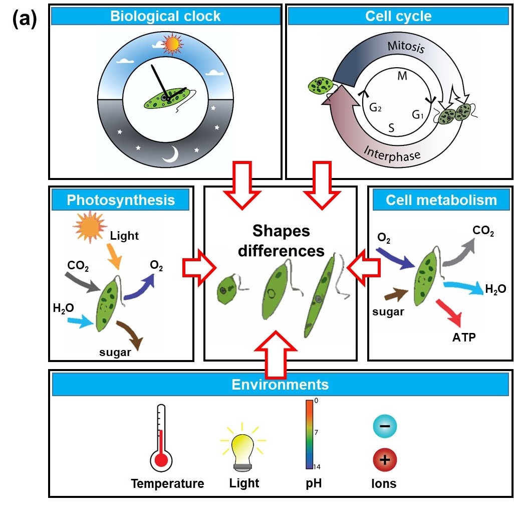
Many aspects of biology have become automated, which allows researchers to monitor the properties of single cells. However, some bottlenecks remain, such as the need to take pictures of cells to detect their shape using expensive equipment and image-recognition software. A better solution would be one that is cheaper and does not require high-speed optics to operate effectively. Now, in a study published in Biosensors and Bioelectronics, a team of scientists led by NAIST has developed a simple and fast method for detecting the shape of cells as they pass through a microfluidic channel based on changes in the electrical impedance.
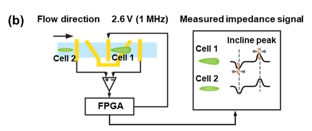
Using a nonlinear electrode layout, the team found that changes in the impedance as the object passed by were sensitive to its shape, but not affected by its trajectory within the microchannel. "We introduced a new metric we call the 'tilt index,' which is zero for symmetric objects, like spherically-shaped cells, but nonzero for elongated cells," lead author Yaxiaer Yalikun explains.
Using a sinusoidal excitation voltage with a frequency of one megahertz, the scientists could detect the resulting signal with a specially built lock-in amplifier. The team tested the system with polystyrene beads with diameters between 10 and 25 microns, as well as living Euglena gracilis cells of varying shapes. E. gracilis is already widely used in biological research and could also become a source of biofuel in the future. "Reducing the cost of automated monitoring is an important step for making new biotechnologies commercially viable," senior author Yoichiroh Hosokawa says. Future versions may be incorporated directly into cell growth systems as a quality control measure, because signs of stress can sometimes manifest as changes in cellular shape.
###
Resource
-
Title: Microscopic impedance cytometry for quantifying single cell shape
-
Authors: Tao Tang, Xun Liu, Ryota Kiya, Yigang Shen, Yapeng Yuan, Tianlong Zhang, Kengo Suzuki, Yo Tanaka, Ming Li, Yoichiroh Hosokawa & Yaxiaer Yalikun
- DOI: 10.1016/j.bios.2021.113521
- Information about Hosokawa's lab can be found at the following website:
https://mswebs.naist.jp/english/courses/list/labo_11.html